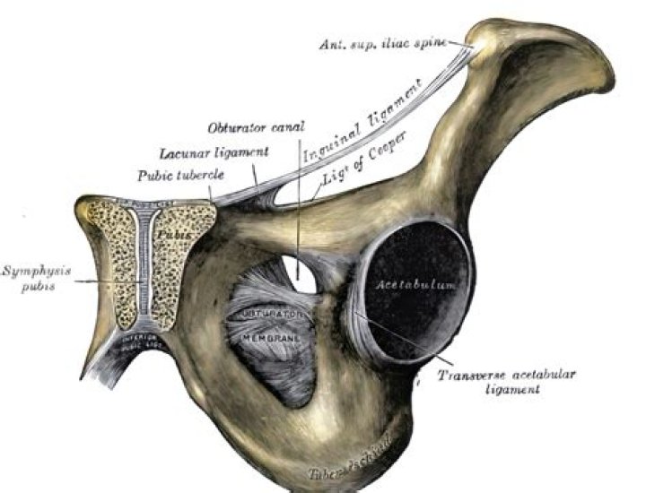
The obturator membrane (latin: membrana obturatoria) is a strong fibrous joint or syndesmosis that fills the obturator foramen of the hip bone. The obturator membrane attaches to the margins of the obturator foramen..
Similarly, you may ask, what is the obturator membrane?
The obturator membrane is a thin fibrous sheet, which almost completely closes the obturator foramen.
Furthermore, where does the obturator artery go? The obturator artery is a branch of the internal iliac artery that passes antero-inferiorly (forwards and downwards) on the lateral wall of the pelvis, to the upper part of the obturator foramen, and, escaping from the pelvic cavity through the obturator canal, it divides into both an anterior and a posterior branch.
Also Know, where is the obturator located?
The internal obturator is situated partly within the lesser pelvis, and partly at the back of the hip-joint. It functions to help laterally rotate femur with hip extension and abduct femur with hip flexion, as well as to steady the femoral head in the acetabulum.
What is the obturator fossa?
The obturator foramen is a large aperture, situated between the ischium and pubis. In the male it is large and of an oval form, its longest diameter slanting obliquely from before backward; in the female it is smaller, and more triangular. Through the canal the obturator vessels and nerve pass out of the pelvis.
Related Question Answers
What does the obturator muscle do?
Obturator internus. Its primary function is to help move the thigh away from the center of the body by rotating it in a sideways direction. When the thigh is flexed, it assists other muscles in moving the thigh outward, away from the midline of the body. It also helps to stabilize the hip joint.What passes through obturator canal?
The obturator canal is a passageway formed in the obturator foramen by part of the obturator membrane. It connects the pelvis to the thigh. The obturator artery, obturator vein, and obturator nerve all travel through the canal.What covers the obturator foramen?
The obturator internus muscle lies on the intrapelvic side of the obturator membrane, a fibrous membrane that covers the obturator foramen. The tendon of the obturator internus muscle traverses the lesser sciatic foramen to insert on the greater trochanter of the femur, facilitating lateral thigh rotation.What muscle passes through the lesser sciatic foramen?
The lesser sciatic foramen has as its boundaries the ischial body anteriorly, the ischial spine and the sacrospinous ligament superiorly and the sacrotuberous ligament posteriorly. The tendon and nerve of obturator internus as well as the pudendal nerve and vessels pass through the foramen.Where is the hip joint found?
The hip joint (see the image below) is a ball-and-socket synovial joint: the ball is the femoral head, and the socket is the acetabulum. The hip joint is the articulation of the pelvis with the femur, which connects the axial skeleton with the lower extremity.What Innervates obturator Externus?
Innervation: Posterior division of obturator nerve innervates most of the adductor magnus; vertical or hamstring portion innervated by tibial nerve. Arterial Supply: Obturator and medial circumflex femoral arteries.What causes obturator pain?
Obturator neuropathy is a difficult clinical problem to evaluate. One possible cause of pain is due to fascial entrapment of the nerve. Symptoms include medial thigh or groin pain, weakness with leg adduction, and sensory loss in the medial thigh of the affected side.What happens if the obturator nerve is damaged?
The obturator nerve can be damaged through injury to the nerve itself or to surrounding muscle tissue. This type of injury can occur during household or car accidents and it can also happen accidentally during abdominal surgery. A damaged obturator nerve can cause pain, numbness, and weakness of the thigh.How do you treat obturator nerve pain?
For anterior obturator nerve entrapment, treatment may consist of electrical stimulation of the adductor and hip flexor muscles, stretching, and massage. These modalities, however, typically have not been successful in resolving this condition if it is not recognized early.What is the obturator sign?
The obturator sign or Cope's obturator test is an indicator of irritation to the obturator internus muscle. The technique for detecting the obturator sign, called the obturator test, is carried out on each leg in succession. The patient lies on her/his back with the hip and knee both flexed at ninety degrees.What muscles does the obturator artery supply?
Supply. The obturator artery supplies the pelvic muscles it crosses, the head of the femur, the muscles of the medial compartment of the thigh and gives a small branch to the knee capsule. The iliac branch supplies the bone and the iliacus muscle. It also has a cutaneous supply to the medial thigh.What does the obturator nerve supply?
The obturator nerve (L2–L4) supplies the pectineus; adductor (longus, brevis, and magnus); gracilis; and external obturator muscles. This nerve controls adduction and rotation of the thigh. A small cutaneous zone on the internal thigh is supplied by sensory fibers.Is the obturator nerve motor or sensory?
The obturator nerve is responsible for the sensory innervation of the skin of the medial aspect of the thigh. The nerve is also responsible for the motor innervation of the adductor muscles of the lower limb (external obturator.What is an obturator hernia?
An obturator hernia is a rare type of hernia of the pelvic floor in which pelvic or abdominal contents protrudes through the obturator foramen. It is characterized by lancilating pain in the medial thigh/obturator distribution, extending to the knee; caused by hernia compression of the obturator nerve.What does the sciatic nerve branch into?
The sciatic nerve is derived from the lumbosacral plexus. Within the posterior thigh, the nerve gives rise to branches to the hamstring muscles and adductor magnus. When the sciatic nerve reaches the apex of the popliteal fossa, it terminates by bifurcating into the tibial and common fibular nerves.What is the crown of death?
Corona mortis, Latin for "crown of death", is a common variant vascular anastomosis between the external iliac artery or deep inferior epigastric artery with the obturator artery.Where does the inferior gluteal artery come from?
The inferior gluteal artery originates as a branch of the anterior division of the internal iliac artery. It approaches the greater sciatic foramen by passing back through the parietal pelvic fascia and between the S1 and S2 nerve roots.What is the corona mortis?
The "corona mortis" is an anatomical variant, an anastomosis between the obturator and the external iliac or inferior epigastric arteries or veins. It is located behind the superior pubic ramus at a variable distance from the symphysis pubis (range 40-96 mm).What is abnormal obturator artery?
Obturator artery (OA) is a branch of anterior division of the internal iliac artery (IIA). In about 20-25% cases, OA arises from the inferior epigastric artery (IEA) instead of the internal iliac artery (EIA) and then it is called an abnormal obturator artery (AOA).