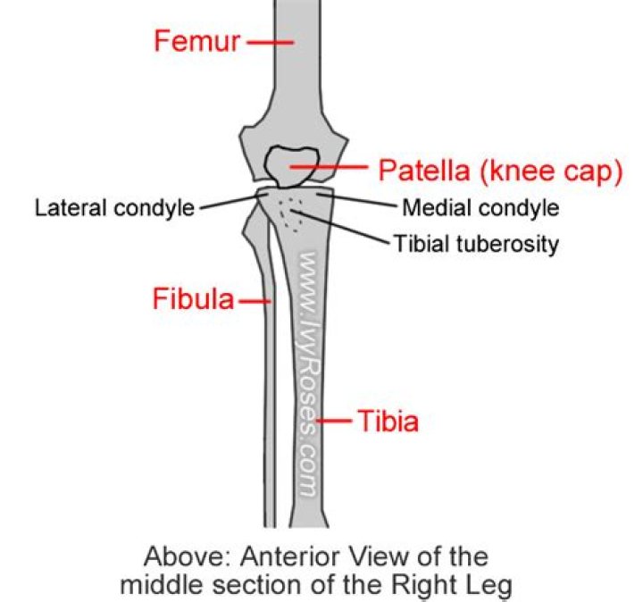
The lateral condyle is the lateral portion of the upper extremity of tibia. It serves as the insertion for the biceps femoris muscle (small slip).
Where are the condyles of the tibia?
Anatomical terms of bone The medial condyle is the medial (or inner) portion of the upper extremity of tibia. It is the site of insertion for the semimembranosus muscle.
What is the lateral condyle of the knee?
The lateral condyle is one of the two projections on the lower extremity of the femur. The other one is the medial condyle. The lateral condyle is the more prominent and is broader both in its front-to-back and transverse diameters.
Where is the lateral tibia located?
Tibialis anteriorLateral surface of tibia, Interosseous membraneSoleusSoleal line, Head of fibula, Posterior border of fibulaWhere is the medial and lateral condyles located?
The medial and lateral condyles form the proximal part of the body of femur, and articulate with the proximal part of tibia to form the femorotibial joint. They are separated by the deep intercondylar fossa, proximally bounded by the horizontal intercondylar line.
What does lateral condyle mean?
Medical Definition of lateral condyle : a condyle on the outer side of the lower extremity of the femur also : a corresponding eminence on the upper part of the tibia that articulates with the lateral condyle of the femur — compare medial condyle.
Is tibia medial or lateral?
The tibia is found on the medial side of the leg next to the fibula and closer to the median plane or centre-line.
Where is the condyle?
A condyle (/ˈkɒndəl/ or /ˈkɒndaɪl/; Latin: condylus, from Greek: kondylos; κόνδυλος knuckle) is the round prominence at the end of a bone, most often part of a joint – an articulation with another bone. It is one of the markings or features of bones, and can refer to: On the femur, in the knee joint: Medial condyle.Where is your tibia bone in your leg?
The tibia, or shin bone, is the larger bone in your lower leg. Beside it, more toward the outside of the leg, is the fibula. The tibia forms part of the knee joint. The ends of the tibia and the fibula both form part of the ankle joint.
What is the bone located on the lateral side of the lower leg?The fibula is the smaller, thinner bone of the lower leg. It is on the lateral side of either leg, meaning it is away from the middle of the body on each side. The head of the fibula attaches to the head of the tibia and does not make up part of the knee joint. The base of the fibula forms part of the outer ankle.
Article first time published onWhat attaches to the medial tibial condyle?
The medial condyle presents posteriorly a deep transverse groove, for the insertion of the tendon of the Semimembranosus. Its medial surface is convex, rough, and prominent; it gives attachment to the tibial collateral ligament.
Where does the tibia meet the knee?
Tibia (Shin Bone) The Tibia meets the Femur at the knee in two areas on which the Femur rides. This area called the Tibial Plateau is divided into a medial (inside of your knee) and a lateral (outside) part.
Where is the lateral malleolus?
The knob on the outside of the ankle, the lateral malleolus, is the end of the fibula, the smaller bone in the lower leg.
What does the lateral condyle of the tibia articulate with?
The lateral and medial condyles articulate with the tibia to form the knee joint. The epicondyles provide attachment for muscles and supporting ligaments of the knee.
Does the tibia have condyles?
The proximal portion of the tibia consists of a medial and lateral condyle, which combine to form the inferior portion of the knee joint. Between the two condyles lies the intercondylar area, which is where the anterior collateral ligament, posterior collateral ligament, and menisci all have attachments.
Where does the tibia end?
The proximal end of the tibia terminates in a broad, flat region called the tibial plateau. The intercondylar eminence runs down the midline of the plateau, separating the medial and lateral condyles of the tibia.
Where does the tibia form?
In humans the tibia forms the lower half of the knee joint above and the inner protuberance of the ankle below. The upper part consists of two fairly flat-topped prominences, or condyles, that articulate with the condyles of the thighbone, or femur, above.
Where is distal tibia?
The distal tibia bears medial and posterior prominences known as the medial malleolus and posterior tibial process, respectively. The medial malleolus is longer than the lateral tibial surface and articulates with the medial surface of the talus to form the medial gutter of the ankle joint.
Does the tibia have Epicondyles?
The proximal end of the tibia is greatly expanded. The two sides of this expansion form the medial condyle of the tibia and the lateral condyle of the tibia. The tibia does not have epicondyles. The top surface of each condyle is smooth and flattened.
What attaches to the lateral condyle of the femur?
The lateral epicondyle of the femur, smaller and less prominent than the medial epicondyle, gives attachment to the fibular collateral ligament of the knee-joint.
How long does it take to heal a lateral condyle fracture?
The most common timeline for this injury is four to six weeks in a long arm cast. Your child will then come back to get the cast removed and have an x-ray. Pins from surgery are removed once the bone has healed and is stable. If screws were used, they may not need to be removed.
What causes pain in shins?
You get shin splints from overloading your leg muscles, tendons or shin bone. Shin splints happen from overuse with too much activity or an increase in training. Most often, the activity is high impact and repetitive exercise of your lower legs. This is why runners, dancers, and gymnasts often get shin splints.
Can you walk on a tibia fracture?
Can you still walk with a fractured tibia? In most cases, the answer is no. Walking after a tibia fracture can make your injury worse and may cause further damage to the surrounding muscles, ligaments and skin. Walking on a fractured tibia is also likely to be extremely painful.
What does a tibia fracture feel like?
Symptoms are very similar to ‘shin splints’ with gradual onset pain on the inside of the shin. Individuals suffering from a tibial stress fracture typically feel an aching or burning (localized) pain somewhere along the bone. Swelling may be present at the fracture site.
Is condyle a bone or cartilage?
About Knee Anatomy Three bones make up the knee joint – the femur, the tibia and the patella. The femur (thigh bone) is the largest bone in the body and extends from the hip to the knee where it ends in structures known as condyles that are covered in cartilage.
What means condyle?
Definition of condyle : an articular prominence of a bone especially : one resembling a pair of knuckles.
What is a condyle?
Condyle – Refers to a large prominence, which often provides structural support to the overlying hyaline cartilage. It bears the brunt of the force exerted from the joint. Examples include the knee joint (hinge joint), formed by the femoral lateral and medial condyles, and the tibial lateral and medial condyles.
Why does the outside of my lower leg hurt?
Shin splints refer to the pain and tenderness along or just behind the large bone in the lower leg. They develop after hard exercise, sports, or repetitive activity. Shin splints cause pain on the front or outside of the shins or on the inside of the lower leg above the ankle.
Why does the side of my calf hurt when I walk?
Claudication is a common condition where pain occurs in the legs with exercise due to a reduction in the circulation. The cause is hardening of the arteries otherwise known as atherosclerosis. The common symptom is of a cramp like pain developing in the calf muscles on walking.
Why does the outside of my calf hurt?
Summary. Calf pain can be caused by injuries to muscles, bones, or tendons as well as infections or conditions that affect blood flow. Your healthcare provider may diagnose the pain using imaging tests or blood tests. Depending on your diagnosis, they may suggest medication, rest, or physical therapy.
Is the lateral or medial condyle larger?
The lateral condyle is slightly wider than the medial condyle at the centre of the intercondylar notch.