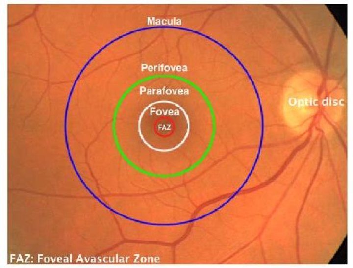
The macula is the pigmented part of the retina located in the very center of the retina. In the center of the macula is the fovea, perhaps the most important part of the eye. The fovea is the area of best visual acuity. It contains a large amount of cones—nerve cells that are photoreceptors with high acuity.
Is fovea and macula the same?
The macula is the center portion of the retina that produces even sharper vision with its rods and cones. The fovea is the pit inside the macula with only cones, so vision can be at its sharpest. While the fovea and the macula have the same objective of providing clear vision, they achieve that goal in different ways.
What is the macula also called the fovea?
The depression in the very center of the macula where eyesight is sharpest. It is also called the fovea centralis. A number of eye problems can affect the fovea and can lead to vision loss if they are not treated.
What is the difference between macula lutea and fovea?
It is the part of the retina that is responsible for sharp, detailed central vision (also called visual acuity). The macula lutea, also called fovea, contains a very high concentration of cones. These are the light-sensitive cells in the retina that give detailed central vision.What is the difference between the macula and the retina?
is that retina is (anatomy) the thin layer of cells at the back of the eyeball where light is converted into neural signals sent to the brain while macula is (anatomy) an oval yellow spot near the center of the retina of the human eye, histologically defined as having two or more layers of ganglion cells, responsible …
Is fovea and yellow spot same?
The yellow spot or macula is an oval yellow spot near the centre of the retina of the human eye. … It is the area of best vision where maximum amount of cone cells are present.It is also known as fovea centralis and Macula Lutea. Most of the sensory cells are present at this spot.
Do all primates have central fovea?
Visual System The fovea is present in the retina of all primates with the exception of prosimians and the nocturnally adapted owl monkey (Aotus sp.). Neurotransmission circuits in the fovea have one-to-one connections between photoreceptors, bipolar cells, and ganglion cells, which allow for maximal acuity [164].
What is dry macular?
Dry macular degeneration is a common eye disorder among people over 50. It causes blurred or reduced central vision, due to thinning of the macula (MAK-u-luh). The macula is the part of the retina responsible for clear vision in your direct line of sight.What is the difference between the fovea and the blind spot?
Visual acuity such as sharpness and detail is greatest at the fovea, while at the blind spot it is insensitive to visual stimulation, it’s the part of the retina that converges to the optic nerve.
What is in the macula?The macula is part of the retina at the back of the eye. It is only about 5mm across but is responsible for our central vision, most of our colour vision and the fine detail of what we see. The macula has a very high concentration of photoreceptor cells – the cells that detect light.
Article first time published onWhat is another name for fovea?
Also called the central fovea or fovea centralis. The word “fovea” is the Latin word for “small pit.” The fovea is literally a small depression (in the retina).
What is the fovea and its function?
The fovea is responsible for sharp central vision (also called foveal vision), which is necessary in humans for activities for which visual detail is of primary importance, such as reading and driving.
What is fovea simple?
Definition of fovea 1 : a small fossa. 2 : a small depression in the center of the macula (see macula sense 2b) that contains only cones and constitutes the area of maximum visual acuity and color discrimination — see eye illustration.
What is the difference between macula and macular?
Macular degeneration deteriorates the retina which is the camera of the eye. AMD impacts the central part of the retina, called the macula, that focuses central vision. The macula is the part of the eye that controls reading, driving, seeing faces and the fine detail of any type of object.
Does the fovea have rods?
The increased density of cones in the fovea is accompanied by a sharp decline in the density of rods. In fact, the central 300 µm of the fovea, called the foveola, is totally rod-free.
What are the 3 layers of the retina?
The cellular layers of the retina are as follows: 1) The pigmented epithelium, which is adjacent to the choroid, absorbs light to reduce back reflection of light onto the retina, 2) the photoreceptor layer contains photosensitive outer segments of rods and cones, 3) the outer nuclear layer contains cell bodies of the …
Do dogs have a macula?
Visual acuity – Dogs do not have a fovea or a macula (area of the retina where there is a high concentration of cones, the day receptors of the retina) and the optic nerve of the dog contains much less nervous fibers than the one of the man.
What is the difference between fovea and Foveola?
In context|anatomy|lang=en terms the difference between fovea and foveola. is that fovea is (anatomy) the retinal fovea, or fovea centralis, responsible for sharp central vision while foveola is (anatomy) the center of the fovea in the macula of the eye, approximately 035 mm in diameter, containing only cone cells.
How does the fovea differ from the rest of the retina?
The retina is a light-sensitive layer at the back of the eye that covers about 65 percent of its interior surface. … In the middle of the retina is a small dimple called the fovea or fovea centralis. It is the center of the eye’s sharpest vision and the location of most color perception.
Why is macula lutea yellow?
Because the macula is yellow in colour it absorbs excess blue and ultraviolet light that enter the eye and acts as a natural sunblock (analogous to sunglasses) for this area of the retina. The yellow color comes from its content of lutein and zeaxanthin, which are yellow xanthophyll carotenoids, derived from the diet.
Which layers of retina is absent in fovea?
Layers or RetinaCommon CharacteristicsFovea/MaculaNerve Fibre LayerAxons of ganglion cellsAbsentInner Limiting MembraneFusion of Muller cell and vitreous membranesPresent, but very thin
What do you mean by rhodopsin?
rhodopsin, also called visual purple, pigment-containing sensory protein that converts light into an electrical signal. Rhodopsin is found in a wide range of organisms, from vertebrates to bacteria.
What is difference between blind spot and yellow spot?
Blind spot is a spot on the retina present at the point of origin of the optic nerve. Yellow spot is a small area on the retina present at the posterior pole of the eye, lateral to the blind spot.
What is the relationship of the fovea to cones in the retina?
In the fovea, there are NO rods… only cones. The cones are also packed closer together here in the fovea than in the rest of the retina. Also, blood vessels and nerve fibers go around the fovea so light has a direct path to the photoreceptors.
Is the blindspot on the fovea of the eye?
The blind spot is located about 15 degrees on the nasal side of the fovea. Healthy humans do not generally notice this lack of visual information since our brain interpolates the blind spot based on surrounding detail, information from the other eye, and the calculation of different images resulting from eye movements.
Does Areds 2 really work?
Results showed that the AREDS2 combination reduced the risk of disease progression by as much as 19 percent and/or of vision loss by 25 percent. In patients with early (Category Two) AMD, the supplements did not slow the disease’s progression to intermediate AMD.
What is difference between wet and dry macular degeneration?
Dry Macular Degeneration. The main difference between wet vs dry macular degeneration is simple: dry macular degeneration is the more common type of eye disease and does less damage to your vision while wet macular degeneration can result in serious vision loss.
What is the best vitamin for dry macular degeneration?
- 500 milligrams (mg) of vitamin C.
- 400 international units (IU) of vitamin E.
- 10 mg of lutein.
- 2 mg of zeaxanthin.
- 80 mg of zinc (as zinc oxide)
- 2 mg of copper (as cupric oxide)
What is macula Cirrus?
The Zeiss Cirrus HD-OCT is a non-invasive technology used for imaging the vitreous and retina — the multi-layered sensory tissue lining the back of the eye. The Optical Coherence Tomography (OCT) scanner provides physicians with an automated, segmented representation of the choroid and retinal layers.
Is the macula a tissue?
A macular pucker is scar tissue that has formed on the eye’s macula, located in the center of the light-sensitive tissue called the retina. The macula provides the sharp, central vision we need for reading, driving, and seeing fine detail.
Where is the macula in the fundus?
A fundus photo, showing the optic disc as a bright area on the right where blood vessels converge. The spot to the left of the centre is the macula.