Dampen a cotton swab with 1 to 2 drops of lens cleaning solution. A cotton swab will help you clean the surface of a concave lens, which curves inward. Be sure to use a lens cleaning solution, or a solvent like acetone or xylol..
Subsequently, one may also ask, why must you use lens paper to clean the objectives and not paper towel?
Kimwipes, paper towels, paper, your fingers, and other materials will damage the lenses. Never use paper or Kimwipes on a lens because they have wood fibers which will scratch a lens! ? These fibers have been removed from lens paper.
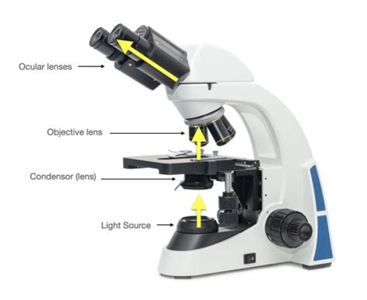
Likewise, what does the lens do on a microscope? The compound microscope has two systems of lenses for greater magnification, 1) the ocular, or eyepiece lens that one looks into and 2) the objective lens, or the lens closest to the object. If your microscope has a mirror, it is used to reflect light from an external light source up through the bottom of the stage.
why is it important to clean the lenses of a microscope?
It is important to clean a microscope of dirt, oil, and stains after use because the lens can easily catch dirt, fingerprints, or culture solution during operation.
What is the safest solvent for cleaning an objective lens?
Acetone is the safest solvent for cleaning an objective lens. Only lint-free, optically safe tissue should be used to wipe off microscope lens. Once the focus is achieved at one magnification, a higher-power objective lens can be rotated into position without fear of striking the slide.
Related Question Answers
When should the lenses be cleaned What is the correct way to clean them?
Do not use any sprays with cleaners. Once blown clean, lightly wipe the lens with Kimwipes or another approved lens cloth. Another good cleaning tissue is Kodak Lens Tissue (available at photo stores) In lieu of a brush, you can use the paper.Do you use alcohol to clean objective lens?
The following liquids can be used for cleaning: Ether:alcohol (80:20 or 70:30, depending on manufactuer): Moisten a cotton swab and clean the optical surfaces, such as the front lens of the eye piece, in a circular manner. Lens paper should not be used, as it may scratch the optical surfaces.What is a coverslip used for?
The main function of the cover slip is to keep solid specimens pressed flat, and liquid samples shaped into a flat layer of even thickness. This is necessary because high-resolution microscopes have a very narrow region within which they focus. The cover glass often has several other functions.What is the proper way to transport a microscope?
When moving your microscope, always carry it with both hands (Figure 1, at left). Grasp the arm with one hand and place the other hand under the base for support. Turn the revolving nosepiece so that the lowest power objective lens is "clicked" into position (This is also the shortest objective lens).What kind of tissue do you use to clean the microscope slides?
Cleaning and Handling Microscope Slides. Samples are placed on thin pieces of glass called microscope slides and covered with thin slivers of glass called coverslips. Many experienced technicians 'huff' on the slide, that is breathe out onto the slide to fog it and then wipe clean with lens tissue.Why is it important to eliminate air bubbles from the slide?
It protects the microscope and prevents the slide from drying out when it's being examined. The coverslip is lowered gently onto the specimen using a mounted needle . It is important that no air bubbles are trapped underneath. Most cells are colourless.WHAT IS lens paper?
Definition of lens paper. : a soft nonabrasive lintless tissue paper used for wiping and wrapping lenses.How do you change the magnification of a microscope?
(Microscopes usually come with a set of objective lenses that can be interchanged to vary the magnification.) You can calculate the total magnifying power of the microscope by multiplying the magnifying powers of the objective lens and the eyepiece (so 10 x 40 = total magnification of 400x).What is the purpose of the iris diaphragm?
Iris Diaphragm controls the amount of light reaching the specimen. It is located above the condenser and below the stage. Most high quality microscopes include an Abbe condenser with an iris diaphragm. Combined, they control both the focus and quantity of light applied to the specimen.HOW IS lens cleaned?
Remove as much dust and dirt as possible from the lens with a blower or soft-bristled brush. Apply a few drops of lens cleaning solution to a lens tissue or cleaning cloth. Using a circular motion, gently remove oil, fingerprints, and grime from the lens surface, working from the center outward.What happens to the amount of light as one increases magnification?
The light intensity decreases as magnification increases. There is a fixed amount of light per area, and when you increase the magnification of an area, you look at a smaller area. So you see less light, and the image appears dimmer. Image brightness is inversely proportional to the magnification squared.When focusing a specimen should you always start with?
ALWAYS use both hands when picking the microscope up and moving it from one place to another. 3. When focusing on a slide, ALWAYS start with either the 4X or 10X objective. Once you have the object in focus, then switch to the next higher power objective.Why is it helpful for a microscope to be Parfocal?
Answer and Explanation: It is helpful for a microscope to be parfocal because the user does not have to adjust the focus when changing the power of magnification.What is the best way to clean immersion oil from the microscope lens?
It can be removed with lens paper dipped in a weak ammonia solution (one dropper full of household ammonia in 1/2 cup water). If you are using a 100x objective with immersion oil, just simply "swipe" the excess oil off the lens with a kimwipe after use.What is total magnification?
Total magnification is when the object being viewed is magnified to its maximum limit.How do objectives magnify an image?
The objective, located closest to the object, relays a real image of the object to the eyepiece. This part of the microscope is needed to produce the base magnification. The eyepiece, located closest to the eye or sensor, projects and magnifies this real image and yields a virtual image of the object.How does a microscope magnify an object?
A microscope is an instrument that is used to magnify small objects. It is through the microscope's lenses that the image of an object can be magnified and observed in detail. A simple light microscope manipulates how light enters the eye using a convex lens, where both sides of the lens are curved outwards.What are the functions of microscope?
First, the purpose of a microscope is to magnify a small object or to magnify the fine details of a larger object in order to examine minute specimens that cannot be seen by the naked eye.What is the function of the stage?
Arm: Structural element that connects the head of the microscope to the base. Stage: The flat platform that supports the slides. Stage clips hold the slides in place. If your microscope has a mechanical stage, the slide is controlled by turning two knobs instead of having to move it manually.